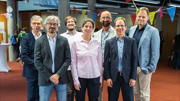
Florian Grüner and Theresa Staufer on their x-ray fluorescence research.“From the Development of New Medications to Measuring Immune Response”
5 October 2022, by Tim Schreiber

Photo: Private
A team from the Department of Physics at Universität Hamburg has managed to measure the uptake of co-enzymes by individual human skill cells. In an interview, Prof. Dr. Florian Grüner and Dr. Theresa Staufer spoke about the methods applied and their potential for further development.
What makes the methods you applied special?
Florian Grüner: Our x-ray fluorescence imaging methods allow high resolution measurements of marked entities, such as individual cells or medications, to determine their distribution spatially as well as over time. There are many varied potential fields of application, ranging from pharmaceutical studies in the development of new medications, through the measuring the immune response in therapy and for chronic inflammatory diseases. Two key characteristics unique to these methods, described to as “multi-tracking” or “multi-scale imaging”, refer to the ability to both track multiple entities at the same time, and also to image them at different scales from whole body scans to individual cells.
How can these methods be further developed?
Theresa Staufer: The two main goals for developing these methods is to create more compact x-ray sources, and also to produce significantly larger detectors for measuring the emission of x-ray signals. Currently, the measurements are conducted using a synchrotron, that is, a big accelerator, and there are not many of those. That means we need much more compact sources, to further develop the methods in laboratories as well as later in clinical applications. The second goal addresses the special x-ray detectors currently available, whose surfaces can only detect a fraction of the spatial angle available, which limits its sensitivity for the time being, until larger-surface solutions can be found.
What are you planning in terms of application for this research?
Florian Grüner: Our working group is working closely with a range of cooperation partners not only from the world of research but also from the clinical field and from industry. In particular, this close collaboration with medical and pharmaceutical research corporations is essential to develop methods that allow us to glean data that we currently cannot capture and thus answer some open questions. In addition, we also have close contact with developers and manufacturers from a diverse range of x-ray sources to make the methods accessible to a broader user-base.
What are some other potential applications?
Theresa Staufer: In addition to application to individual cells, the methods may make a significant contribution to medications, for example, in cancer therapies and monitoring immune responses in cell therapy, as well as the early detection of tumors. The methods can also assist with online-diagnostics in therapy, and quantify the immune system response after radiation.
How important is collaboration in the research center of Hamburg?
Florian Grüner: The research center of Hamburg has the unique advantage that there are many experts under one roof. We can only push these methods forward with close cooperation between the University Medical Center Hamburg-Eppendorf, researchers from the fields of organic chemistry, nanosciences, and physics from both Universität Hamburg and DESY. This is because the methods cannot be conducted without suitable markers and x-ray sources.


