2017 Nobel Prize in Chemistry: structural biologist Prof. Dr. Kay Grünewald on the importance of cryo-electron microscopy
6 October 2017, by Maike Nicolai

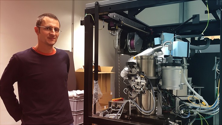
Photo: CSSB
This year's Nobel Prize in Chemistry goes to three pioneers of cryo-electron microscopy, a path-breaking technology enabling us to examine native biological samples using high resolution at low temperatures. Structural biologist Prof. Dr. Kay Grünewald researches in this field and explains to us why this technology is such a huge breakthrough for biology.
Why is the development of cryo-electron microscopy such a significant scientific step?
Cryo-elctron microscopy (cryo-EM) allows us to analyze in 3D the individual macromolecules directly in their native watery environment and their functional interaction. This also closes a gap in biological structural determination.
Previously, specimens were typically prepared by chemical fixation, dehydration, staining, and embedding in resins. These methods also create artefacts. The cryo-preparation introduced by Jacques Dubochet’s team, however, leaves the specimen in its watery environment while preserving its structural integrity all the way down to atomic level.
About 4 years ago there was a breakthrough in resolution with the introduction of direct electron detectors. The Prizewinner Richard Henderson and his group were among the leaders in this field. The detectors make proteins—down to their amino acid side chains—visible at atomic level, for example. We can now use this structural information to decode and understand fundamental biological functions.
What technical challenge does cryo-electron microscopy pose?
We need to master three technical challenges:
1) Specimens must be frozen very quickly. The goal is to avoid the development of crystalline ice and to embed them in glass-like ice. This process is called “vitrification.”
2) Specimens prepared thus are cooled with liquid nitrogen at roughly 180 C and analyzed using very low electron radiation in the electron microscope. The latter is important to avoid damaging the structure of the biological specimen. Highly sensitive cameras—nowadays generally direct electron detectors—create images of the specimen.
3) Finally, using sophisticated methods of analysis—Prizewinner Joachim Frank and his group did the groundwork here—several of these images are calculated, visualized, and analyzed as three-dimensional biological macromolecules with the aid of computer clusters.
What exactly are you researching at the Centre for Structural Systems Biology (CSSB) using cryo-electron microscopy and what insights do you hope to gain?
At CSSB we are primarily investigating molecular interactions between pathogens and their host cells. In my working group, these pathogens are generally viruses. The goal is to better understand important steps in a virus’ entry into the cell, the assembling of the virus, and its morphogenesis. This also helps us find approaches to intervention. We focus on herpes, adeno-, and retroviruses.
We also use viruses as markers to work on basic questions in cell biology. To do so, we use cryo-electron microscopy; one of two main modalities of cryo-EM allows us to analyze macromolecules directly in their native cellular environment.
This way we can grasp important cellular mechanisms such as intracellular transport and membrane infusion. This involves combining information from the cryo-EM using an integrative structural biology approach and findings from other fields such as biochemistry, X-ray crystallography, or mass spectrometry.
Good to know:
In 2017 Prof. Dr. Kay Grünewald received funds to the tune of €15.6 million für Universität Hamburg to establish a cryo-electron microscopy facility in the new building of the Centre for Structural Systems Biology (CSSB) on the Bahrenfeld Campus.


